
-
by Tamer
When discussing X-ray image quality, mAs and kVp are often the first parameters that come to mind. They are essential for achieving appropriate exposure and diagnostic image quality, but they are only part of the equation.
Whether you work with digital radiography (DR), general X-ray systems, mammography or DEXA (also called DXA) technology, understanding the relationship between imaging parameters, equipment performance and clinical technique is essential.
Excellent image quality comes from understanding how every component of the imaging process works together.
Here are eight factors that can make a meaningful difference to your images.
1. Source-to-Image Distance (SID)
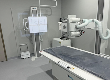
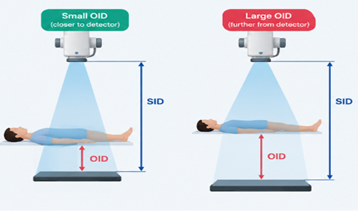
Source-to-image distance (SID) affects beam intensity at the detector and influences magnification and geometric sharpness. Because beam intensity falls with the square of the distance, increasing SID generally reduces magnification and improves sharpness, although maintaining receptor exposure may require adjustments to exposure factors.
Choosing the appropriate SID for each examination helps achieve consistent image quality while supporting efficient X-ray workflows.
2. Object-to-Image Distance (OID)
Object-to-image distance (OID) is often overlooked, yet it has a direct impact on image geometry.
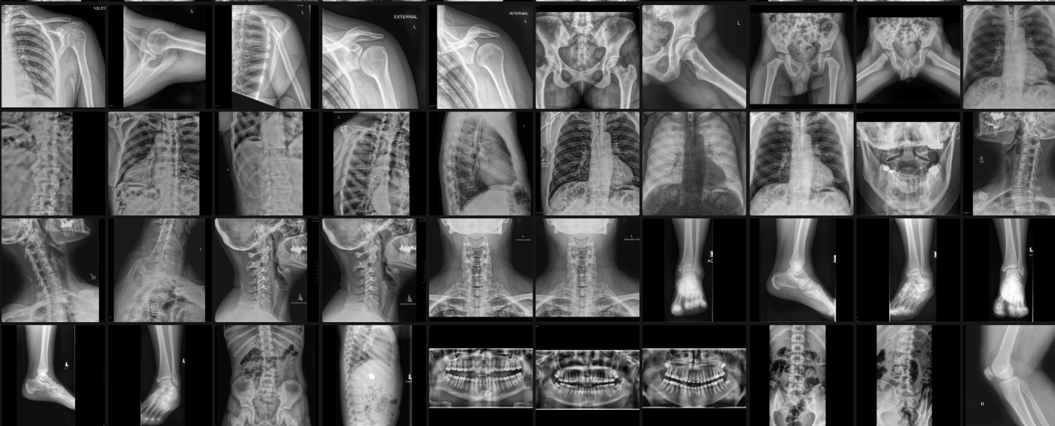
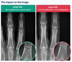
As the distance between the patient and detector increases, magnification and geometric unsharpness generally increase. Keeping OID as small as practical can help improve recorded detail, particularly when imaging small structures. The diagram and radiographs below show the difference.
3. Patient Positioning
Even with advanced digital radiography equipment, poor positioning can compromise diagnostic value.
Rotation, incorrect centring, movement and inappropriate patient alignment can obscure anatomy or create unwanted distortion. In specialised examinations such as mammography, accurate positioning is particularly important for demonstrating the required anatomy.
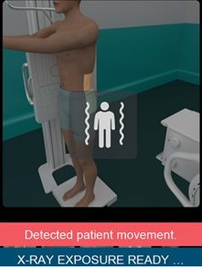
Careful positioning helps ensure diagnostic consistency and can reduce the need for repeat exposures. Technology can help too: the new DRGEM collimator includes a built-in camera that detects patient movement and alerts the operator before the X-ray is taken.
4. Collimation
Collimation is much more than simply restricting the radiation field.
Appropriate collimation reduces the volume of tissue irradiated and can reduce scatter reaching the detector, improving image contrast. It also supports radiation protection by limiting unnecessary exposure, a priority highlighted in ARPANSA’s Safety Guide for Radiation Protection in Diagnostic and Interventional Radiology. Accurate collimation is an important consideration across general X-ray imaging and other radiographic examinations.
5. Focal Spot Selection
The focal spot size influences spatial resolution.
A smaller focal spot can provide better recorded detail by reducing geometric unsharpness. However, its use may be limited by tube loading and exposure requirements.
Selecting the appropriate focal spot for the examination means balancing image detail, exposure time and equipment limitations.
6. Detector Performance and Digital Radiography
Modern digital radiography (DR) systems offer advanced image acquisition and processing capabilities, but not all detectors perform identically.
Detector pixel size, spatial resolution, detective quantum efficiency (DQE) and noise characteristics all influence the final image. The scintillator also matters, so see our comparison of CsI and GOS flat panel detectors.
Manufacturers such as DRGEM develop digital X-ray systems for diagnostic imaging environments, and HEDI Tech supplies DRGEM radiography systems to Australian healthcare providers. However, achieving excellent results depends on more than the equipment alone. Appropriate exposure technique, patient positioning, detector performance and system configuration all contribute to the final image.
Still working with an analogue or computed radiography (CR) room? A DR upgrade can bring digital detector performance to your existing system, and our CR vs DR article explains the differences.
7. Grid Selection and Exposure Index
Grids can improve image contrast by reducing the amount of scatter reaching the detector, particularly when imaging thicker anatomy. However, they also absorb some primary radiation and may require increased exposure. For more detail, read our guide oscillating vs non-oscillating grids.
The exposure index (EI) provides useful feedback about detector exposure, but it is not a direct measurement of patient dose or image quality. Standard definitions of EI are set out in IEC 62494-1 and explained in the AAPM Task Group 116 report.
Understanding the target exposure index and its relationship to the imaging system helps support consistent image quality and appropriate exposure optimisation.
8. Post-Processing
Post-processing plays an important role in achieving optimal image quality by refining how the acquired image is displayed. Understanding parameters such as contrast, brightness, edge enhancement and noise reduction helps optimise image presentation for different anatomical structures and clinical requirements.
However, post-processing cannot recover anatomical detail that was never captured during acquisition. A good understanding of these tools allows radiographers to make informed adjustments, improve image consistency and avoid excessive enhancement that may obscure important diagnostic details.
Improving Image Quality Across X-ray, Mammography and DEXA
Although the specific requirements vary between modalities, the underlying principle remains the same: equipment performance and clinical technique must work together.
In general X-ray and DR imaging, geometry, collimation, positioning and detector characteristics are important considerations. In mammography, positioning, compression, image resolution and specialised system performance are critical to producing diagnostically useful images.
DEXA systems have different imaging objectives, focusing on bone mineral density and, where supported, body composition analysis. Here, consistent patient positioning, appropriate scanning protocols, calibration and quality control are essential for reliable measurements. Learn more about DEXA bone densitometer systems or read about the expanding role of DEXA.
Quality Medical Imaging Solutions in Australia
At HEDI Tech, we help Australian healthcare providers bridge the gap between quality and cost, delivering reliable, high-performance medical imaging solutions across general X-ray, mammography, DEXA, OPG and CBCT, backed by trusted technology partners including DRGEM, Genoray, OsteoSys, RAY and Vatech.
Explore our full range of medical imaging systems, read what to consider when buying an X-ray machine, or contact our support for a free consultation.
